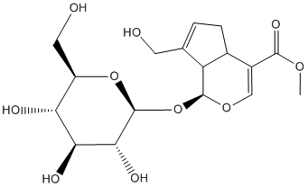
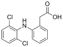
VL is a major public health importance in Indian subcontinent with more than 90% of the world’s cases and affects the poorest population mainly in rural areas. Emergence of resistance against pentavalent antimony, the mainstay of treatment, in north-eastern India and the toxicity, availability and affordability of second-line drugs leaves the situation more complicated. Human VL is characterized by a marked humoral response and impaired Sipeimine cell-mediated immunity,  associated with an inability to control infection. During VL infection, impairment of Diacerein nitric oxide generation and IL-12 production from macrophages occurs whereas the disease promoting cytokines TGF-b and IL-10 are enhanced. Still, it has been demonstrated that the ability to mount a pro-inflammatory response is critical for control and eventual resolution of infection and that the Th1 cytokines IL-2, IL-12 and IFN-c are essential for response. Because of the lack of effective and low-cost treatments and the irreversibility of tissue damage during infection, considerable attention has been focused towards vaccine development. Recent research on leishmaniasis has been focused towards determining strategies that specifically stimulate protective immune responses in the absence of those that may cause pathology and/or interfere with protection. Thus, leishmanial antigens that predominantly stimulate Th1 responses in patient cells or rodents infected with the parasite have been accepted as ��potential protective antigens’and therefore promising vaccine candidates. Earlier studies in our laboratory, by using classical activity based fractionation and sub-fractionation of the soluble proteins from clinical isolate of L. donovani promastigote, led to the identification of a potent sub-fraction ranging from 89.9 to 97.1 kDa which induced Th1 type cellular responses in cured Leishmania patients and hamsters along with significant prophylactic efficacy in hamsters. Further proteomic characterization of this subfraction led to the identification of 18 Th1 stimulatory proteins and among them Enolase and Fructose bisphosphate Aldolase, the vital proteins belonging to the glycolytic pathway, were also present. Aldolase is a central glycolytic enzyme in carbohydrate metabolism, catalyzing the cleavage of fructose 1,6-bisphosphate into two triose sugars, glyceraldehyde 3-phosphate and dihydroacetone phosphate whereas Enolase is known to catalyse the reversible dehydration of D-2-phosphoglycerate to phosphoenolpyruvate in both glycolysis and gluconeogenesis. During the past decade glycolytic enzymes have emerged as vaccine targets/candidates in many different organisms. This has been attributed because of their moonlighting functions associated along with their classical functions in glycolysis. In Fasciola hepatica and Schistosoma mansoni, recombinant FBA was confirmed to provide significant protection for experimentally infected animals. Further, FBA was immuno-screened with sera from patients infected with Onchocerca volvulus, S. mansoni, Streptococcus pneumonia and Plasmodium falciparum. It is also considered as a potential diagnostic antigen in case of human giardiasis. A unique b-3-FBA conjugate was developed by Xin et al, wherein protection against C. albicans infection was uniquely acquired through immunity against the carbohydrate and the FBA peptide. In Leishmania, apart from its glycolytic function Enolase also localizes on the surface and binds plasminogen which contributes to the virulence of the parasite. Therefore, these are vital for energy production that is necessary for parasite activities and survival.
Monthly Archives: May 2019
Extracellular sulfate can diffuse freely through the outer membrane into the periplasmic region and bind to the VFT2 region of BvgS
In previous comparative genomic studies we showed that ptxP1 and ptxP3 strains have different SNPs in a number of virulence-associated genes, differences in pseudogenes, as well as differences in gene content. A recent transcriptional comparison of ptxP1 and ptxP3 strains under non-modulating Yunaconitine conditions indicated that multiple virulence-associated genes are expressed at slightly higher levels in ptxP3 strains as compared to ptxP1 strains. Here, we compared sulfate-dependent expression profiles between these strains, with the rationale that Diperodon sulfate affects the expression of all major B. pertussis virulence factors. This approach identified several additional differences between the ptxP1 and ptxP3 strains, including sulfate-dependent differences in expression levels of a number of important virulence genes and a different sensitivity for sulfate-mediated regulation. Conceivably, the two are interconnected. The most pronounced phenotypic difference revealed by microarray analysis was that ptxP1 and ptxP3 strains respond differently to sulfate mediated-regulation. Based on genome annotations and BLAST searches we identified 30 genes that are likely involved in sulfate metabolism, nine of which were differentially regulated between the ptxP1 and ptxP3 strain. The sulfate genes included genes involved in uptake and metabolism of sulfate, cysteine, methionine and taurine. Interestingly, taurine is one of the most abundant sources of sulfate in the host, comprising  0.1% of the total human body weight. In general, the sulfate genes were expressed at higher levels by the ptxP3 strain under low sulfate conditions compared to the ptxP1 strain. For instance, the sbp gene, which facilitates transport of external sulfate into the cell, showed a 9-fold higher level of gene expression in the ptxP3 strain. Furthermore, nine other sulfate genes were expressed at two- to four-fold higher level in the ptxP3 strain under low sulfate conditions, although not all values reached statistical significance. The increased expression of sulfate genes in the ptxP3 strain may be explained by the observation that the positive master regulator for cys gene expression, cysB, was more highly expressed in ptxP3 strains than ptxP1 strains under low and medium sulfate conditions. It is tempting to speculate that the expression profile of these genes contributes to the reduced sensitivity of ptxP3 strains to sulfate-mediated suppression of known Bvg-regulated genes as shown in this work. For instance, we found that ptxP3 strains in particular, and to a lesser extent ptxP1 strains, expressed higher levels of Ptx in the presence of 5 mM sulfate, whilst ptx genes have been described to be suppressed under this condition. This suggests that the protein expression pattern of Ptx in ptxP3 strains more accurately reflects a Bvgi-phase protein than the classical Bvg+ profile. Recently, it was described that the BvgS sensor molecule is active by default and is only inhibited when sulfate or other negative modulators bind to the Venus Flytrap 2 region in the periplasmic domain of this sensor molecule. Whilst the concentration of free sulfate in the respiratory tract is low, infection may increase sulfate concentration locally, e.g. through desulfation of sulfated host proteins, potentially through pertussis proteins containing a sulfatase domain of which ORF BP3136 was expressed above the expression minima in both strains. Interestingly, the wcbQ gene encoding a capsular polysaccharide biosynthesis protein with a sulfatase domain, was expressed exclusively and at 4.5 fold higher levels in the ptxP1 strain under low and medium sulfate conditions, as compared to the ptxP3 strain.
Such diversity of physiological actions makes these receptors increasingly important as drug targets
In most cases, obtaining a sufficient amount of a purified membrane protein for biophysical studies requires a heterologous expression system. In this respect, production of recombinant GPCRs relies on bacterial, yeast, eukaryotic cells or cell-free methods, each method presenting its own advantages. The E. coli host, utilized in the present work, lacks endogenous Gproteins, necessary for GPCR signaling, but has great potential due to its simplicity, growth at high density, possibility to scale up, safety, and low cost. It can also employ the power of bacterial genetics with different strains and expression vectors. Most bacteria lack post-translational modifications, such as glycosylation, which may be critical for GPCRs function in eukaryotic cells, but also have the advantage of yielding homogeneous samples for structural investigations. Finally, bacteria allow for easy stable isotope labeling for protein NMR studies. Recombinant GPCRs expressed in E. coli can be obtained by producing the protein in a membrane-integrated form followed by detergent solubilization. Although limited in part by differences, such as presence of cholesterol, between the bacterial and the eukaryotic cell membrane, the E. coli expression LOUREIRIN-B system allows the GPCR to fold and assemble in an environment relatively similar to the native one. Mammalian GPCRs produced in E.  coli membranes include b2-adrenergic receptor, serotonin 5-HT1A receptor, neurotensin receptor, Nk-2 receptor, M1 muscarinic acetylcholine receptor, M2 muscarinic acetylcholine receptor, adenosine A2A receptor, CB1-CB2 cannabinoid receptors and CCR5-CCR3-CXCR4CX3CR1 chemokine receptors. A recent study has described the expression of numerous GPCRs in E. coli membranes. Despite its promise, GPCR expression in E. coli membrane is highly unpredictable, i.e. dependent upon the particular GPCR, and often results in a very low yield. Bacterial overexpression of GPCRs can also result in incorrect folding and protein aggregation in the form of cytoplasmic inclusion bodies. These have limited toxicity to the bacterial host and may protect the protein from lysis. Therefore, as an alternative to membrane insertion, a second GPCR production strategy in bacteria consists of directing recombinant product into inclusion bodies, followed by protein denaturation and refolding. However, it is to be taken into account that GPCR refolding might present challenges and that not all GPCRs expressed in E. coli produce inclusion bodies. Several class B1 ECD-1 3D-structures, including CRFR ECD1s, both in isolation or in complex with protein ligands have been determined. Importantly, the class B1 structures of the trans-membrane domains of CRFR1a and of glucagon Ginsenoside-F2 receptor have been recently determined. In order to promote functional analyses and structural studies we have undertaken the characterization of recombinant human CRFR1a and mouse CRFR2b produced in bacterial membranes. Using a series of expression plasmids, we verified the impact of different strains, culture media and protein SPs. Detergents were screened to optimize the solubilization of the receptors from the bacterial membranes. The functionality of the receptors expressed in E. coli intact cells and corresponding membrane fractions was assessed by radioreceptor assays using selected CRF family ligands. In the E. coli host, several factors may affect both the yield of recombinant membrane protein and its distribution between membrane and inclusion bodies such as transcriptional promoter, translational initiation efficiency, codon usage, mRNA secondary structure and stability, protein translocation efficiency, protein stability, and its level of toxicity to the bacterial host.
The association of GPCRs with biological membranes makes their structural analysis especially challenging
Migration involves a wide array of cellular changes including alterations in cellular structure by regulation of cytoskeleton dynamics and expression of adhesion molecules. Therefore, changes in actin structure have the ability to directly affect cell migration. ROS are also known to actively participate in each of the  above events. Since MWCNT significantly induces the generation of ROS and actin expression, it is of interest to evaluate the effects of MWCNT on cell migration. The cell scratch assay results clearly show that MWCNT exposure can increase cell migration, while pretreatment of NAC abolishes this effect, which are consistent with other studies regarding the effects of MWCNT on cell migration. It has been reported that MWCNT could trigger inflammatory response in mice or cell lines, and the increased ROS production is at least partially responsible for it, although contradictive results also exist. In our study, several proteins involved in inflammation and infection processes, such as Annexin A1, Complement component 1 Q subcomponentbinding protein, Poly-binding protein 2 are indentified. Additionally, we also find an elevated ROS production. Therefore, it is of interest to know whether MWCNT could influence the inflammatory response in A549 cells. Among the Th1/Th2 cytokines examined, only IL-6 secretion is affected by MWCNT treatment. Thus, the IL-6mediated inflammatory response might be a major target of MWCNT. Several of the other Chloroquine Phosphate identified proteins are also of interest for further study. 14-3-3e is the most conserved member of the 14-3-3 family, and is involved in a wide range of physiological processes. For example, homocysteine induces apoptosis of rat hippocampal neurons by inhibiting 14-3-3e expression, and downregulation of 14-3-3e causes partial meiotic resumption of the mouse oocyte. Moreover, a significant reduction of 14-3-3e protein expression is Oxysophocarpine detected in gastric cancer. Given the prevalence of reduced 14-3-3e expression in such processes, the importance of reduced 14-3-3e expression after MWCNT exposure merits further investigation. Another interesting protein is Hsp27, a member of the small heat shock protein family, which protects against apoptotic cell death induced by a variety of stimuli including elevated temperature, heavy metals, oxidative stress and cytotoxic agents. Our initial prediction is that under stress conditions, such as MWCNT exposure, HSP expression would have been induced for its protective function, however, we find instead that HSP27 expression is decreased. The physiological significance of such changes requires further clarification. With approximately 800 different genes, G-protein-coupled receptors constitute the largest family of human integral membrane proteins. By transducing a wide array of extracellular stimuli into intracellular signals, they participate in many fundamental biological processes. In addition, because of their important role in pathological processes and cell membrane localization, they represent very prominent drug targets. Structurally, GPCRs are characterized by an extracellular Nterminus, followed by seven transmembrane segments, which are connected by intracellular and extracellular loops, with the Cterminus located inside the cell. Based on their sequence similarity, they are divided in five classes. GPCR stimulants range from photons, odorants, and small molecules to large peptide and protein hormones. Upon ligand binding, GPCRs act as guanine nucleotide exchange factors for G proteins, which in turn modulate cellular enzymes or channels.
This was ascribed to another, pathological pathway of fibrin formation initiated by free iron
Moderate iron loading is also known to accelerate thrombus formation after arterial injury, to increase vascular oxidative stress, and to impair vasoreactivity. Furthermore, iron-induced vascular dysfunction may contribute to the increased incidence of ischemic cardiovascular events that have been associated with chronic iron overload. Poorly liganded iron is the main culprit, and plays a fundamental role in the development of pathology, while copper dysregulation may also be of significance. One of the main medical complications of Alprostadil hemochromatosis is that uncontrolled iron leads to tissue damage derived from free radical toxicity caused by the excessive levels of this metal. Previously, we have shown that when ferric iron is added to whole blood taken from healthy individuals, the red blood cell morphology is changed. We have also seen that in diabetes RBCs are elongated and twist around fibrin fibers. The possible hydroxyl radical formation, due to excess iron, or the excess iron itself, may therefore, as can many drugs, change red blood cell and fibrin fiber ultrastructure, as both RBCs and coagulation factors are exposed to Ascomycin particularly high serum ferritin and/or iron that may ultimately cause hydroxyl radicals to be produced in the serum. In view of the above, we here investigate the RBC morphology in blood from individuals with hemochromatosis, in the presence or absence of thrombin, and also study fibrin fiber morphology. To make the logic behind this study and its mechanistic hypotheses the clearer, and following the precepts of Wong, we now include a descriptive Figure 1 that sets out the structure of the manuscript. We also add iron-chelating and other compounds to determine any stabilizing effect of these substances on both RBC ultrastructure and fibrin fiber morphology. We observe remarkable difference in morphology between RBCs from hemochromatosis and hyperferritinemic individuals relative to those from normal controls, with certain kinds of iron chelator and hydroxyl radical scavenger being able to return the aberrant morphology to nearnormal states. It is unknown as to whether this aberrant morphology of itself contributes to disease pathology, but the present findings would possibly open the debate about the desirability of using blood donations from HH individuals. Excess iron levels are associated with Alzheimer’s disease, Parkinson’s disease, Huntington’s disease, Friedreich’s ataxia and other neurological disorders, cancer, Fanconi anemia, stroke, heart disease, diabetes and ageing. One of the hereditary diseases necessarily associated with iron overload is hemochromatosis, where iron overload causes oxidative stress that ultimately damages organs. An unanswered question remains as to what extent, and by what mechanisms, hereditary iron overload conditions may contribute to the clinical manifestation of the conditions listed above. We have recently shown that high added iron may impact on the coagulation profile and RBC ultrastructure. Previously, we have documented that ferric ions can activate nonenzymatic blood coagulation, resulting in the formation of fibrinlike dense matted deposits demonstrable by SEM. Azizova and co-workers also noted that iron causes oxidative modification of thrombin. RBCs also change morphology under elevated iron levels. Further, it is also known that hemoglobin content in HH is raised ; however, the ultrastructure of fibrin fibers and RBCs in blood in HH seem not to have been investigated previously. Currently, we know that iron may change fibrin fiber shape and  packaging, when added at physiological levels. We have also shown that in diabetes, if elevated iron levels are present, changes in fibrin fibers as well as RBC structure occur.