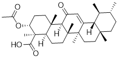
Therefore, the high IAPP concentrations found in the present study are unlikely to result from the presence of autoantibodies against IAPP. Detection of high IAPP plasma 14alpha-hydroxy-Sprengerinin-C concentration in 25 of the 224 tested samples was unexpected, since these patients had developed clinical T1D. Increased IAPP plasma concentration and amyloid formation has been shown to be associated with type 2 diabetes, but we found high IAPP levels in children and adolescents with classic T1D, which develops when less than 10% of the beta cell function remains. The co-storage of IAPP and insulin in the secretory granules normally results in a simultaneous release of the two polypeptide hormones and they are expressed in parallel in the beta cells in response to glucose. Thus a decrease in insulin secretion would be expected to be accompanied by a decrease in IAPP release. We found no correlation between high IAPP levels and high BMI. Neither did we find any correlation to severity of diabetes or other islet hormones such as proinsulin or glucagon, reflecting islet cell function. It has been reported that beta cells depleted of extracellular calcium secrete minimal amounts of insulin both at 1.67 and 16.7 mmol/L glucose in contrast to IAPP where the glucose stimulated secretion was not altered by calcium depletion. The secretion through the regulated pathway is blocked in the absence of calcium while the constitutive pathway would remain unaffected. IAPP may therefore be secreted both through the regulated and the constitutive pathway and secretion of IAPP and insulin may not always be coupled. This may be of importance as insulin in a strong inhibitor of the aggregation of IAPP into toxic assemblies. One could ask if IAPP produced outside the pancreas could be responsible for the high plasma concentrations seen in our T1D patients. In man, pancreas has been shown to be the main source for IAPP but there are also IAPP secreting cells in the pyloric Ergosterol antrum. To what degree this rather limited number of cells could be the source and contribute to the plasma levels of IAPP is unknown, but most likely the cell number  is not sufficient. In summary, while high IAPP expression and amyloid formation so far has been seen in type 2 diabetes, we show that a quite substantial group of children and adolescents with classic T1D have high IAPP concentrations in plasma at the clinical onset of disease. The analysed samples in our study represent the onset of the clinical disease, even though the autoimmune disease process may have lasted for long time. High concentration of IAPP increases the risk for cell toxicity and amyloid formation, particularly in the absence of insulin. Therefore we speculate that the marked increased IAPP concentrations may constitute a risk factor for beta cell destruction also in T1D. It is possible that the group with high IAPP plasma concentration could represent a subgroup within T1D subjects. Further studies of the possible importance of IAPP for development of T1D are obviously highly needed. Actins are highly conserved proteins ubiquitously expressed in eukaryotic cells. In vertebrates, three main groups of actin isoforms have been identified, i.e., alpha, beta and gamma. While the alpha actins are found in muscle tissues and are a major constituent of the contractile apparatus, beta and gamma actins coexist.
Any binding to NIAPP IAPP and IAPPC with plasma from high or low IAPP groups
Leave a reply